Figure 2.

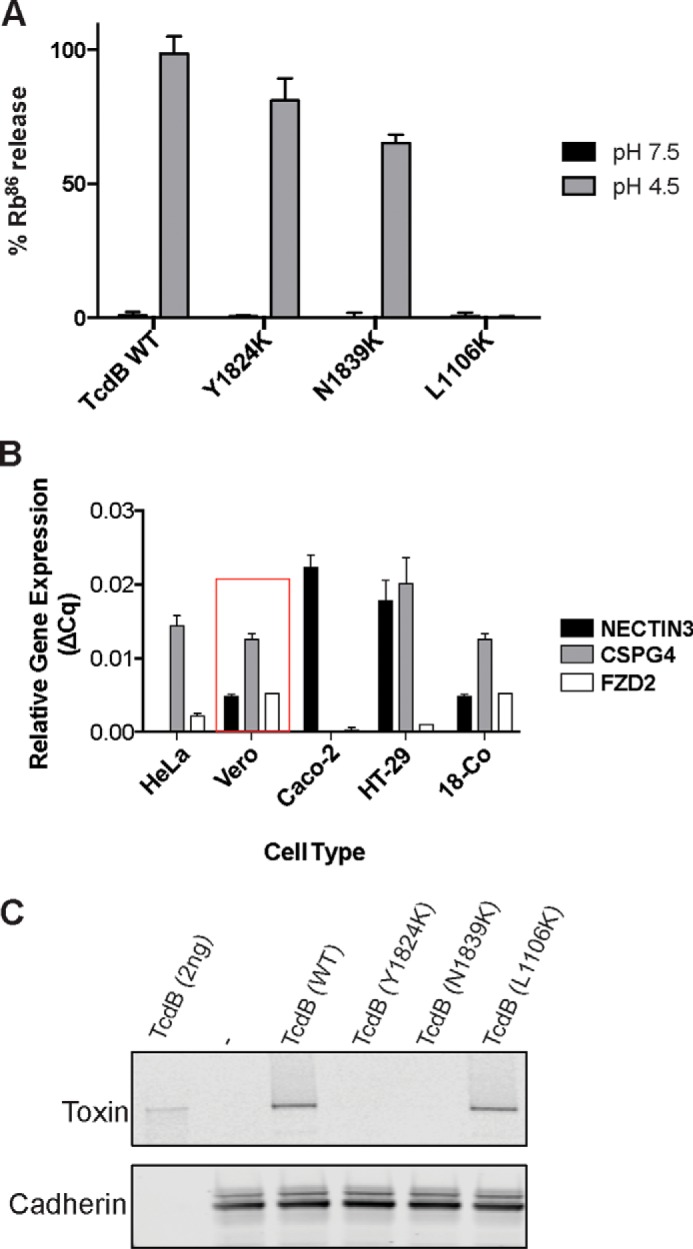
Characterization of defective purified TcdB mutants Y1824K and N1839K. A, pore formation of purified mutant toxins was tested on CHO cells preloaded with 86Rb+. Pore formation was induced by acidification of the external medium (black bars, control, pH 7.5; gray bars, pH 4.5) n = 4. B, relative gene expression of TcdB receptors. The relative gene expression of identified TcdB receptors NECTIN3, CSPG4, and FZDs on different mammalian cell lines was assessed using a ΔΔCq method determined from quantitative PCR data (31) using β-actin, ACTB, as the reference gene (n = 2). Vero cells that were used in the cell viability assay are highlighted in the red box. C, cell-surface binding of defective mutant toxins to target cells. Shown is an immunoblot analysis of TcdB wild type and mutants bound to the Vero cell surface at 4 °C. The cells were exposed to 200 ng/ml TcdB for 30 min before being lysed for immunoblot analysis. Membrane-bound proteins were detected by anti-TcdB antibody and rabbit anti-cadherin antibody as loading control.
