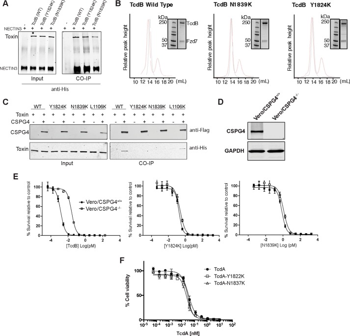
Figure 3.
Characterization of receptor binding of defective mutants. A, association of ectodomain of NECTIN3 with TcdB wild type and mutants, assayed by co-immunoprecipitation (CO-IP) analysis. His-tagged TcdB (3 μg) was incubated with 5 μg of the His-tagged NECTIN3 ectodomain at 4 °C for 16 h. TcdB and NECTIN3 interaction was immunoprecipitated and detected using anti-His antibody. B, interaction of Fzd7 with TcdB by gel filtration assay. Purified Fzd7-CRD mVenus was mixed with TcdB at 1:5 molar ratios of TcdB:Fzd7 and incubated at room temperature for 30 min and run on a Superose 6 Increase 10/300 GL column. Elution peaks of only TcdB (black) and the complex with both TcdB and Fzd7 (red) were monitored by absorbance at 280 nm, and the complex was visualized on SDS-PAGE. Note that all samples were run on the same gel; however, to simplify the figure, the relevant portion of the gel was shown with the corresponding column elution profile. Accordingly, the molecular weight marker was reused in all panels. C, binding of CSPG4 to TcdB wild type and mutants, assayed by co-immunoprecipitation analysis. TcdB (3 μg) was mixed incubated with 150 μg of cell lysate from HEK293 cells overexpressing FLAG-tagged CSPG4 at 4 °C for 2 h. The complex was immunoprecipitated using anti-FLAG magnetic beads and visualized by anti-FLAG (CSPG4) and anti-His (TcdB) antibodies. D, immunoblot analysis of the wild type (CSPG4+/+) and CSPG4 knock-out (CSPG4−/−) Vero cell lysates for expression of CSPG4 and GAPDH (control). E, sensitivity of wild type (CSPG4+/+) and CSPG4−/− Vero cells toward TcdB wild type and mutants. Indicated Vero cells were exposed to TcdB (wild type, Y1824K, and N1839K) and subjected to cell survival using the SRB assay. F, toxicity of TcdA wild type and mutants (Y1822K and N1837K) on CHO cells, measured by Prestoblue fluorescence assay as described.

