Figure 2.
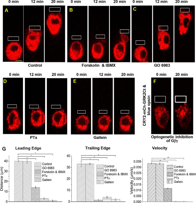
Effect of cAMP/PKA, DAG/PKC, and Gαi-GPCR pathways on RAW cell migration. RAW cells in glass-bottomed dishes expressing blue opsin-mCh and treated with 50 μm 11-cis-retinal were optically stimulated using localized OA (white box) under the following experimental conditions. A, control cells showed complete cell migration with synchronized movement of the LE and the TE. B, 1 μm forskolin and isobutylmethylxanthine prevented the TE retraction, whereas the LE activities remained unperturbed, demonstrating the effect of increased cAMP on cell migration. C, treatment with 10 μm GO6983, a potent PKC inhibitor, did not affect RAW cell migration, suggesting that the DAG/PKC pathway may not have a significant effect on migration. D, incubation of cell with PTx (0.05 μg/ml) for 12 h completely suppressed cell migration. PTx induces ADP-ribosylation and prevents G protein heterotrimer dissociation. E, RAW cells treated with the Gβγ inhibitor, gallein (10 μm, 30 min, 37 °C) also failed to migrate and showed neither LE nor TE activities. F, images of RAW cells transfected with blue opsin GFP, CRY2-mCh-GRK2Ct, and CIBN-CaaX. Local OA resulted in recruitment of CRY2-mCh-GRK2Ct to the PM, near the region of OA. Here, blue opsin activation resulted in release of free Gβγ subunits. However, the recruited CRY2-mCh-GRK2Ct is expected to sequester free Gβγ, preventing activation of its effectors. Even after 20 min of OA, initiation of cell migration was not observed. G, histograms show the extent of movement and migration velocities of the peripheries of the LE and the TE of control and pharmacologically perturbed RAW cells in A–E. *, p < 0.001. (n = 3–10 cells). Scale bar, 10 μm. Error bars, S.E.

