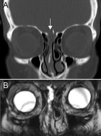
Figure 1.

Representative images from a patient with an anterior sCSF leak. (A) Coronal CT showing a defect in the right cribriform plate (arrow). (B) Coronal T2 MRI showing the resulting meningocele through the right cribriform plate into the nasal cavity.
