


Figure 1.
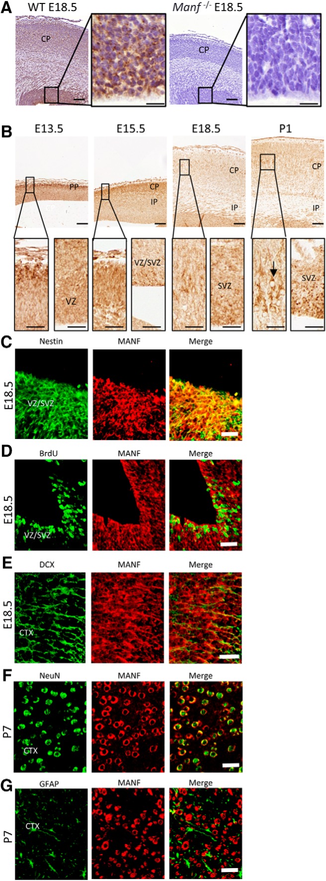
MANF expression in the developing cerebral cortex. A, Coronal cortical sections from E18.5 WT and Manf-/- brains stained with MANF antibody. The insets show photomicrographs of the VZ/SVZ in higher magnification. B, In the E13.5 and E15.5 brain MANF is predominantly localized in cells of the PP/CP and modestly localized in the VZ/SVZ. By E18.5, strong MANF expression is seen in the VZ/SVZ and CP (inset). At P1, MANF is highly expressed in the VZ/SVZ and middle layers of the CP (inset). At E18.5, VZ/SVZ was double-stained for Nestin (green) and MANF (red) antibodies (C) and for BrdU (green) and MANF (red) antibodies (D). E, Section of cerebral cortex from E18.5 mice double-stained with DCX (green) and MANF (red) antibodies. F, Section of cerebral cortex from P7 mice double-stained with NeuN (green) and MANF (red) antibodies. G, Section of cerebral cortex from P7 mice double-stained with GFAP (green) and MANF (red) antibodies; scale bars: 100 µm (A, B), 20 µm (A, B inset), 50 µm (C–G). PP, preplate; CP, cerebral plate; IP, intermediate plate.