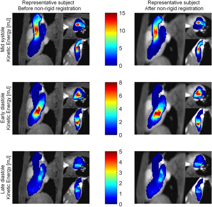
Figure 9.

MIP of the kinetic energy (mJ) of one representative subject in three orientations: three‐chamber view, short‐axis view, and two‐chamber view. Top: mid‐systole, middle: early diastole, and bottom: late diastole. Left: before nonrigid registration, right: after nonrigid registration to the average heart.
