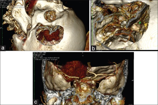
Figure 7.

Planning a simulation of a craniotomy of the left anterior clinoid meningioma with the use of CT angiography DICOM data. (For details, see Video 8.) (a) Simulation of pterional craniotomy. Because of their rich vascularity, the meningiomas are presented very clearly on the reconstructions. (b) After adjustment of the levels of the image (WL/WW & CLUT) hyperostosis is presented where the main blood supply of the tumor is. (c) Note the displacement of the internal carotid artery from the tumor
