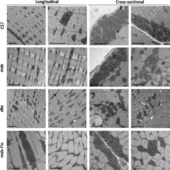
Fig. 2.

Transmission electron micrographs of tibialis anterior muscles from C57BL/10, mdx, dko and mdx-Fiona mice. Representative images of longitudinal sections and cross-sections of tibialis anterior muscles from C57BL/10 (C57), mdx, dko and mdx-Fiona (mdx-Fio) mice (n = 3/group). All scale bars = 2 μm, magnification varies
