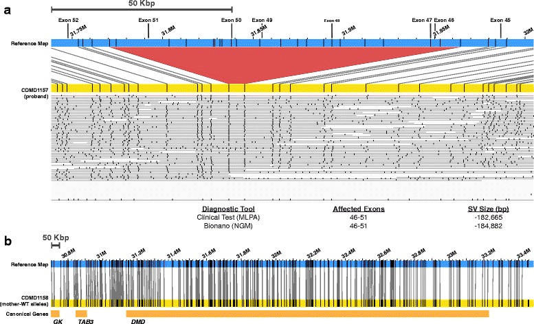
Fig. 6.

NGM identified a hemizygous multi-exon deletion in a DMD patient that was not present in the biological mother. a, b Top: visual representation of the sample allele in yellow (a patient; b mother) compared to the reference (blue). The de novo deletion is shown in red. a Middle: the lines below the patient’s contig represent the long molecules used to construct the sample map. Bottom: Ref-seq locations on the X chromosome indicating possible size of the deletion based on MPLA and size identified using the NGM platform. b Bottom: location of Ref-Seq genes in the X chromosome within the shown region. Maps were generated using Nt.BspQI nicking endonuclease
