
Fig. 7.
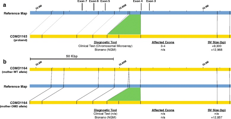
NGM identified a 13-Kbp insertion in a DMD patient and his biological mother. a Top: visual representation of the insertion (green) between the reference (blue) and patient (yellow) maps. Bottom: insertion size identified in the proband by chromosomal microarray and by NGM platform. b Top: the normal wild type allele of the mother (yellow) can be seen above reference (blue) where all nicking sites align to reference map. This is in contrary to the second allele of the mother (yellow) containing the insertion shown below the reference (blue) map. Maps were generated using Nt.BspQI nicking endonuclease