
Fig. 1.
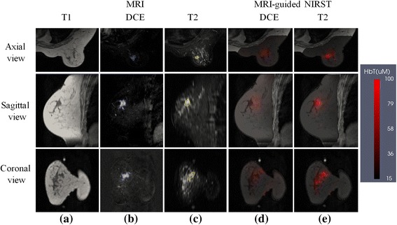
Example patient with a malignant lesion. a T1 MRI; b DCE MRI; c T2 MRI; d,e reconstructed HbT images with DCE-guided and T2-guided methods, respectively. Reconstructed images are overlaid on the T1 MRI cross-section. DCE dynamic contrast-enhanced, HbT total hemoglobin, MRI magnetic resonance imaging, NIRST near-infrared spectral tomography