Figure 2.

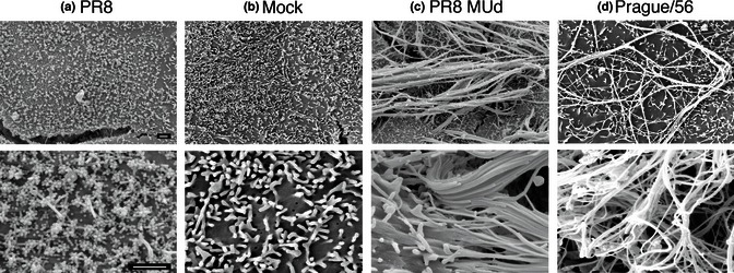
Filament formation by the prototype H7N7 equine influenza A virus. MDCK cells were infected with the indicated viruses at an MOI of 3, fixed at 14 h p.i. and imaged by SEM. Scale bars: 1 μm.

Filament formation by the prototype H7N7 equine influenza A virus. MDCK cells were infected with the indicated viruses at an MOI of 3, fixed at 14 h p.i. and imaged by SEM. Scale bars: 1 μm.