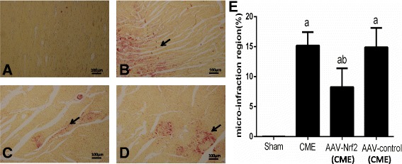
Fig. 1.

Microinfarction detected with HBFP staining (×100), bar = 100 μm. The normal myocardial staining was yellow, while the stain of ischemic myocardium was reddish. Arrows reveal microinfarction focus. a Sham; b CME; c AAV-Nrf2(CME); d AAV-control (CME). e The percentage of microinfarction area. AAV-Nrf2(CME): rats injected Nrf2 undergone CME operation; AAV-control (CME): rats injected AAV-control undergone CME operation; CME: coronary microembolization; Nrf 2: nuclear factor erythroid 2-like; AAV: adeno-associated virus. Data is shown as Mean ± SD. a: P < 0.05 contrasted with sham; b: P < 0.05 contrasted with CME or AAV control (CME)
