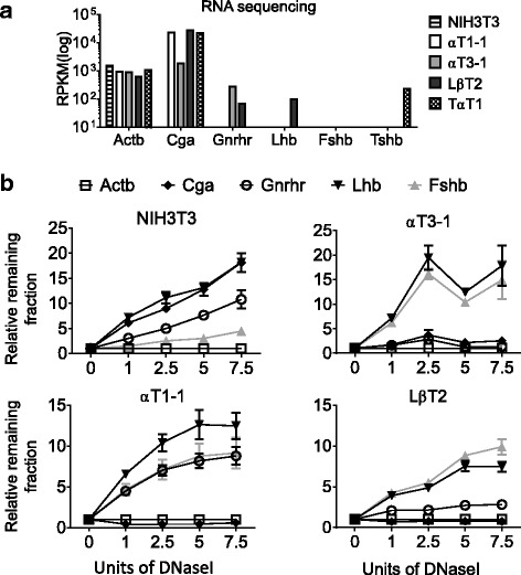
Fig. 1.

Chromatin accessibility of gonadotrope-specific genes during development. a Expression levels of gonadotrope genes in αT1–1, αT3–1, LβT2, TαT1, and NIH3T3 cell lines (n = 2). b DNaseI sensitivity assays in NIH3T3, αT1–1, αT3–1, and LβT2 cells. DNA from nuclei was treated with increasing concentrations of DNaseI and analyzed by qRT-PCR with primers specific to regulatory elements (Table 1). Amplicon quantities were normalized to the active Actb gene, and qRT-PCR data are presented as the mean fraction of DNA remaining relative to Actb ± SEM
