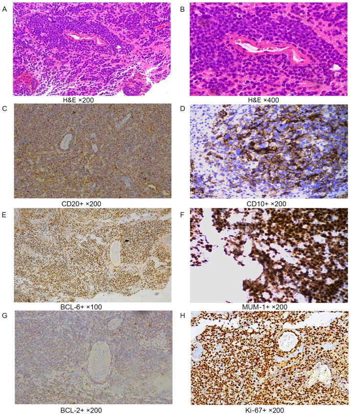
Figure 1.
Immunohistochemical labeling. (A and B) H&E staining. In tumor cells with the diffuse distribution, the size of nuclei were 2 times greater than of normal lymphocytes. (A) Magnification, ×200. (B) Magnification, ×400. (C) CD20 cell membrane staining performed using the EnVision method. Magnification, ×200. (D) CD10 cell membrane staining performed using the EnVision method. Magnification, ×200. (E) BCL-6, nuclei staining, EnVision method, ×100. (F) MUM-1 nuclei staining performed using the EnVision method. Magnification, ×200. (G) BCL-2 cytoplasmic staining performed using the EnVision method. Magnification, ×200. (H) Ki-67 nuclei staining performed using the EnVision method. Magnification, ×200. H&E, hematoxylin and eosin; CD, cluster of differentiation; BCL, B cell lymphoma; MUM-1, multiple myeloma-1.

