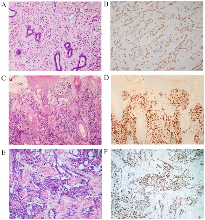
Figure 2.
Immunohistochemical features of angiosarcoma. Tumor cells stained positively for the cluster of differentiation 31 antibody with (A) hematoxylin and eosin staining; and (B) immunohistochemical staining, original magnification, ×100. Nuclear erythroblast transformation-specific-related gene staining with (C) hematoxylin and eosin staining; and (D) immunohistochemical staining, original magnification, ×100, and Friend leukemia virus integration 1 staining with (E) hematoxylin and eosin staining; and (F) immunohistochemical staining, original magnification, ×200 were positive in tumor cells.

