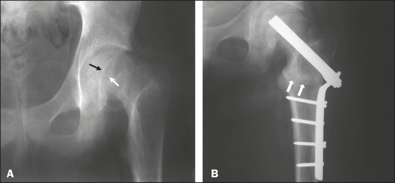
Figure 1.
A: Initial radiograph of the left hip, in an anteroposterior view, showing signs of epiphysiolysis, with inferomedial displacement of the femoral head and an open physis (arrows). B: Radiograph of the left hip, in an anteroposterior view, after valgus intertrochanteric osteotomy; note the positioning of the osteotomy (arrows), fixed with plates and screws.

