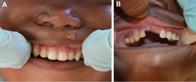
Figure 1.

(A) Mild to moderate acute necrotising gingivitis (ANG) in a 3-year-old girl from the village of Droum. In the region of the upper-right incisors and the canine, the gingiva is oedematous and the interdental papillae have been lost due to ulceration. Soft and hard bacterial deposits are visible. (B) Advanced ANG in a 4-year-old girl from the village of Guidimouni. There are marked signs of ulceration and necrosis of the gingiva, with greyish pseudo-membranes and disappearance of the gingival papillae. The crowns of the teeth are covered with large amounts of soft and hard bacterial deposits.
