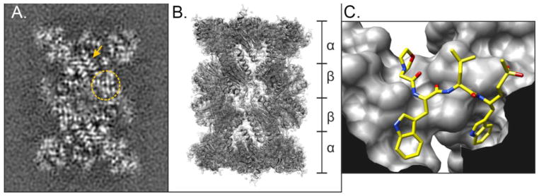
Fig. 2. Visualizing the Plasmodium falciparum 20S core particle.
(A) Cryo-EM image of the P. falciparum 20S core particle (CP). Image shows regions where β-sheet structures (circle) and α-helices (arrow) can be visualized. (B) Fitted model of the 20S core particle based on the cryo-EM images, denoting the stacked α and β rings. (C) Ball-and-stick representation of the parasite-specific peptide vinyl sulfone inhibitor Mu-WLW-vs [18] bound in the β2 active site of the 20S CP. The image shows the large S1 and S3 pockets that can accommodate the two tryptophan residues that do not fit in the human active site.

