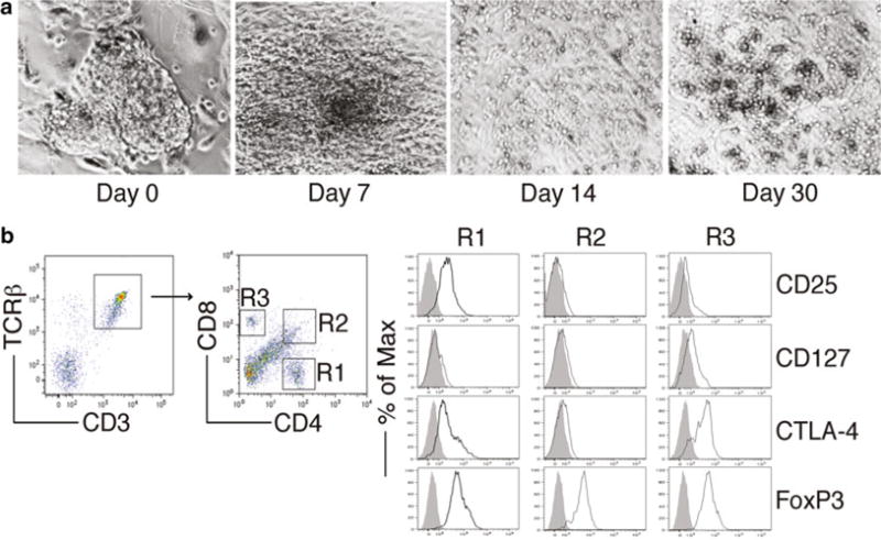
Fig. 2.

FoxP3-transduced iPS cells were cocultured on OP9-DL1/M I-Ab cells in the presence of murine recombinant Flt3L and IL-7. (a) Morphology of Treg cell differentiation on days 0, 7, 14, and 30. (b) Flow cytometric analysis for the protein expression of iPS cell-derived cells on day 30. CD3+ TCRαβ+ cells were gated as indicated, and analyzed for the expression of CD4 and CD8, with CD25, CD127, CTLA-4, and FoxP3 expression shown for cells gated as CD4+ cells (R1), CD4+CD8+ cells (R2), and CD8+ cells (R3) (dark lines, shaded areas indicate isotype controls)
