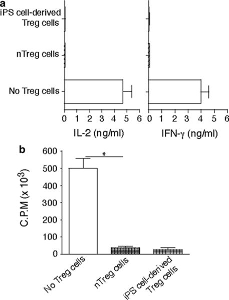
Fig. 4.

Murine iPS cell-derived Treg cells were cocultured with naive CD4+ T cells from C57BL/6 mice (Tregs/T cells = 1:1) in the presence of anti-CD3/anti-CD28 mAbs for 2 days. A group of CD4+ T cells stimulated with CD4+ CD25+ Treg cells from C57BL/6 mice as the positive control and a group of CD4+ T cells stimulated without Treg cells as the negative control. Cytokine production was analyzed by ELISA, and proliferation was determined by [3H] thymidine incorporation. (a) IL-2 and IFN-γ. (b) Thymidine incorporation during the last 12 h. *P< 0.05, one-way AN OVA tests
