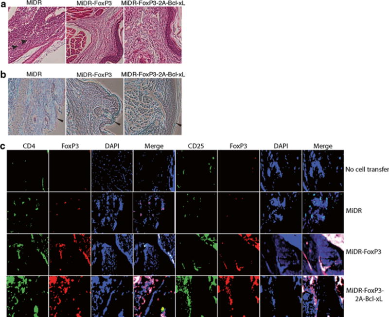
Fig. 5.

Murine iPS cells were transduced with retroviral constructs: vector (MiDR), FoxP3 (MiDR-FoxP3), or FoxP3 with Bcl-xL (MiDR-Bcl-xL-2A-FoxP3), and cocultured on OP9-DL1/MIAb cells. On day 7, DsRed-positive (DsRed+) T cells were sorted and prepared for adoptive cell transfer. Collagen-induced arthritis (CIA) was induced in male C57BL/6 mice by one (day 0) intradermal immunization at two sites in the base and slightly above of the tail with chicken type II collagen in complete Freund’s adjuvant. On day 15 after the immunization, mice received transduced DsRed+ cells (2.5 × 10⁶/mouse). On day 60 of immunization, hind foot paws were amputated, fixed, and decalcified. The tissues were embedded in paraffin, sectioned, and stained. (a) Hematoxylin and eosin (HE) staining. (b) Safranin O–Fast Green staining. Infiltrations of polymorphonuclear (PMN) cells (arrow heads) in HE staining and massive destruction of cartilage (leftward arrow heads) in Safranin O–Fast Green staining are indicated. (c) Immunofluorescent staining. There were iPS cell-derived Treg cells (red) infiltrating in joints from mice receiving iPS cell-derived Treg cells, but not in tissues from mice receiving vector control-transduced iPS cells or without cell transfer (green: CD4 or CD25, red. FoxP3, blue: DAPI).
