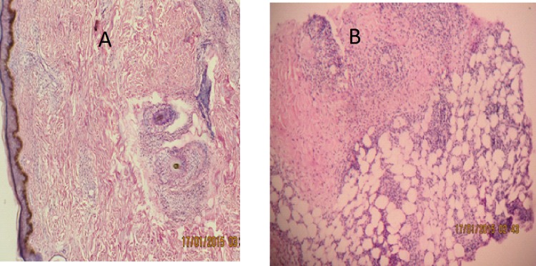
Fig 4.
H &E stained skin biopsies: A. Histopathology of LL lesion without reaction: reticular dermal infiltration of lymphocytes, flat epidermis and foamy histiocytes.B. Histopathology of EN lesion: flat granular PMN infiltration with perivascular lymphocytic infiltration and lobar panniculitis. H & E staining x40.

