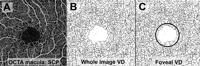
Figure 2.
Processing of macular OCTA scans for measurement of VD. (A) Raw image of macular OCTA scan of the SCP. (B) The raw image was made binary and skeletonized for calculation of whole image vessel density. (C) A 1 × 1 mm circular area centered on the fovea was selected for measurement of foveal VD.

