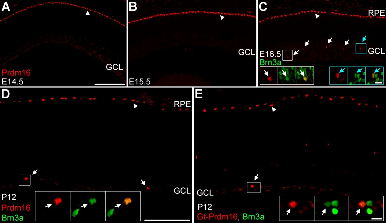
Figure 1.
Prdm16 is made by the RPE and some ganglion cells during development. (A–D) Sheep anti-Prdm16 immunostaining (red) of retinal sections. (A, B) At E14.5 (A) and E15.5 (B), Prdm16 nuclear staining is seen in the RPE (arrowheads), but not within the GCL or other areas of the retina. (C) At E16.5, Prdm16 stains the RPE (arrowhead) and some cells in the GCL (arrows). All Prdm16+ cells in the GCL coexpress the ganglion cell marker Brn3a (green) (arrows, insets). (D) At P12, Prdm16 expression marks the RPE (arrowhead) and a small number of Brn3a+ ganglion cells (arrows, inset). (E) Goat anti-Prdm16 immunostaining (red) at P12 marks the RPE (arrowhead) and a subset of Brn3a+ ganglion cells (arrow, insets). The sheep and goat antibodies have equivalent staining patterns. Weak staining is seen in the ganglion cell layer with both antibodies and may represent either spurious signal or low level expression within other cells. Scale bar: 100 μm for (A–C), and 100 μm for (D, E). Scale bars for insets in (C) through (E) are 10 μm.

