Figure 4.
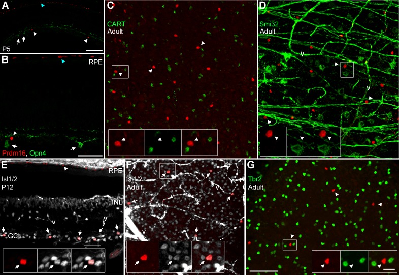
Prdm16+ ganglion cells express Isl1/2, but not other subtype-restricted markers. Sections and flatmounts immunostained with Prdm16 (red). (A, B) P5 retinal sections stained with antibodies against melanopsin (green; Opn4). Melanopsin+ RGCs (arrows) do not coexpress Prdm16 (arrowheads). Blue arrowheads mark the RPE. (C) Adult retinal flatmount stained for CART (green). Prdm16+ ganglion cells do not coexpress CART (arrowheads, insets). (D) Adult flatmount stained for nonphosphorylated neurofilament H (Smi32; green). Prdm16+ cells (arrowheads, insets) do not coexpress Smi32. The fibrous green signal orthogonal to the axons represents nonspecific staining of blood vessels (v). (E) P12 retinal section stained for Isl1/2 (gray). All Prdm16+ cells in the GCL coexpress Isl1/2 (arrows, insets). Arrowhead marks the RPE. (F) Adult flatmount retina stained for Isl1/2 (gray). All Prdm16+ cells coexpress Isl1/2 (arrows, insets). The intense signal in (E) and (F) represents nonspecific staining of vascular structures (v). (G) Adult flatmount stained for Tbr2. No Prdm16+ cells (arrowheads, insets) coexpress Tbr2. Scale bars: 100 μm for (A) and 50 μm for (B), (C) through (G). Scale bar for insets is 10 μm.

