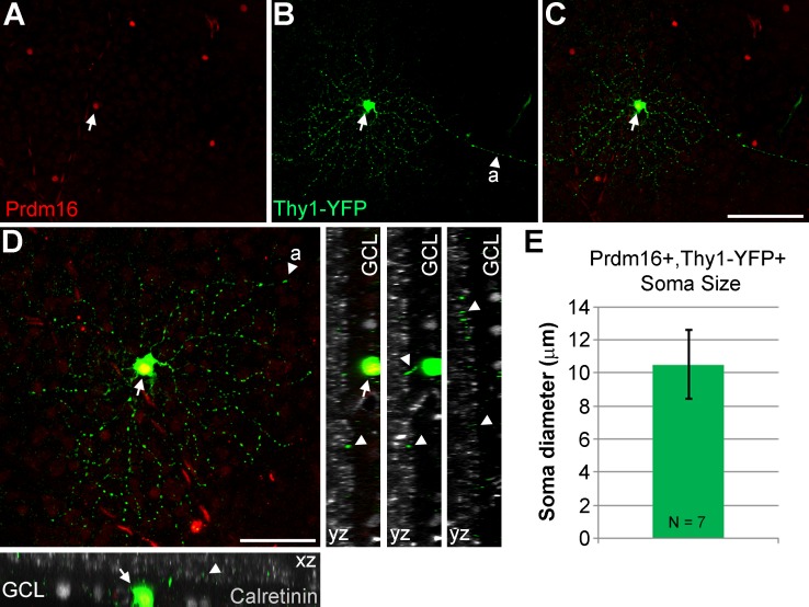
Figure 5.
Morphologic characteristics of Prdm16+ ganglion cells. (A–D) A Thy1-YFP-H transgenic mouse retinal flatmount stained with antibodies to GFP (green); Prdm16 (red); and calretinin (gray). One round Prdm16+ nucleus coexpresses YFP (arrows) and its axon is conspicuous ([A], arrowheads). This cell is located about two-thirds of the way to the retinal periphery (left). (D) A maximum intensity projection image of “C,” rotated and magnified to highlight the dendritic arbor. The dendritic field was 189.6 μm in diameter and circular in shape with few dendrites crossing one another. Dendrites tend to branch sharply. XZ and YZ views of the cell with calretinin to mark the substrata of the inner plexiform layer. The dendrite staining (arrowheads) is localized between the inner and middle calretinin band, within the ON portion of the inner plexiform layer. Scale bars: 100 μm for (A) through (C), and 50 μm for (D). (E) Plot of the average soma diameter measured from seven Prdm16+/Thy1-YFP+ cells. The error bar represents the SD of the seven cell somas measured.

