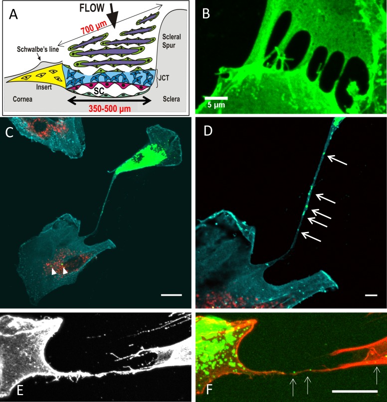
Figure 1.
(A) Schematic of a cross-section of the TM, with the regions and approximate dimensions of the tissue indicated. TM cells (green); corneoscleral beams (purple); the JCT region (blue); the inner wall cells of Schlemm's canal (pink); the putative stem cell insert region (yellow); and Schlemm's canal. (B) TM cells immunostained with CD44 to label the cell membrane. Filopodia from neighboring cells appear to touch and fuse to form a tube. Scale bar: 5 μm. (C) In coculture experiments, DiO-labeled vesicles (green) were present in a DiD-labeled (red) cell (arrowheads). Cyan: CD44; scale bar: 20 μm. (D) At higher magnification, DiO-labeled vesicles are clearly visible (arrows) within a long cell process connecting the two cells. Scale bar: 5 μm. (E) CD44 immunostaining (gray) of a TNT connecting two TM cells. (F) DiO-labeled vesicles (green; arrows) are clearly visible within the SiR-actin-labeled cell process (red). Scale bar: 20 μm.

