Figure 2.
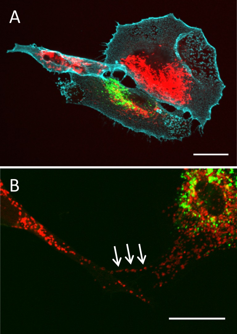
Transfer of mitochondria via TNTs. TM cells were labeled separately with dye (Thermo Fisher Scientific) to stain mitochondria (red) or the fluorescent dye, DiO (green), then mixed 1:1. (A) After overnight incubation, mitochondria (red) were observed in a DiO-labeled cell (green). Cyan: CD44 immunostaining. (B) A higher magnification image of labeled mitochondria (red; Thermo Fisher Scientific) in a DiO-labeled cell (green). Arrows point to mitochondria in a TNT. Scale bar: 20 μm.

