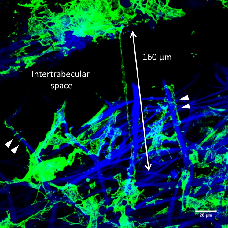
Figure 4.
Frontal section of human TM tissue stained with AlexaFluor phalloidin (green) to label F-actin. A long, actin-rich cell process (approximately 160 μm) bridges an intertrabecular space between corneoscleral beams (double-headed arrow). Other F-actin-rich processes are tightly wrapped around the underlying collagen beams (arrowheads). Blue: DAPI staining and autofluorescence of collagenous TM beams. Scale bar: 20 μm.

