
Figure 3.
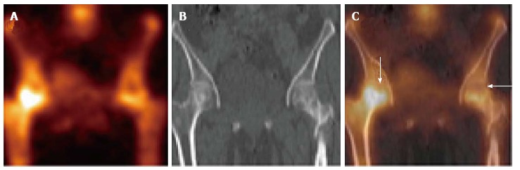
Coronal single photon emission computed tomography (A), coronal computed tomography (B) and coronal fused single photon emission computed tomography/computed tomography images (C) of the patient mentioned in Figure 2 localizes the photopenic defects to head of bilateral femora. The lucent areas with surrounding sclerosis in both femoral heads on low dose computed tomography (CT) component of single photon emission computed tomography/CT (B) increase the diagnostic confidence and specificity.