
Figure 4.
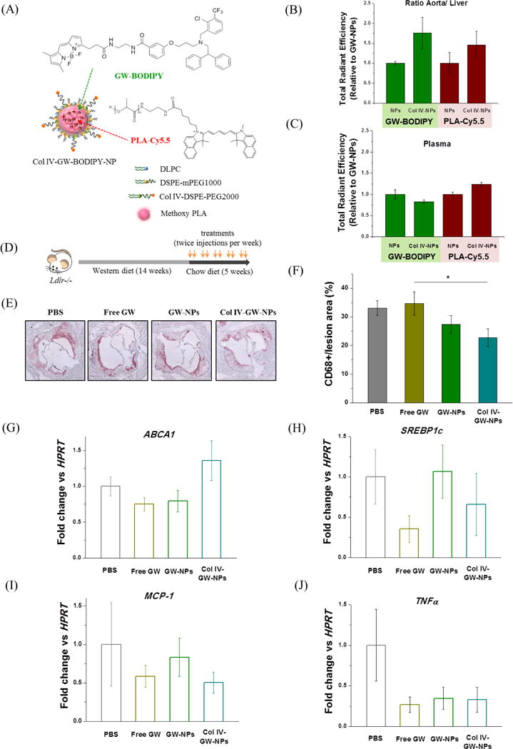
Plaque-targeting abilities and therapeutic efficacies of GW-NPs and Col IV-GW-NPs in the Ldlr−/− mouse model of atherosclerosis. (A) Schematic of the fluorescently labeled Col IV-targeting NP. Fluorescently labeled GW (GW-BODIPY) was encapsulated in the polymeric core of the NP labeled with PLA-Cy5.5 to obtain Col IV-GW-BODIPY-NP. The Ldlr−/− mice (n=2/group) fed a western diet for 14 weeks were injected intravenously with 8 mg of GW/kg body weight in a single dose of fluorescently labeled NPs 4 hours prior to tissue collection. (B) Relative accumulation of GW-BODIPY and PLA-Cy5.5 in the aorta in comparison to the liver. (C) Relative amounts of GW-BODIPY and PLA-Cy5.5 in plasma. (D) Ldlr−/− mice were fed on a western diet for 14 weeks then switched to a chow diet for another 5 weeks. While on the chow diet, the mice were intravenously injected twice a week with 8 mg/kg of body weight (based on the amount of free GW) of free GW, GW-NPs or Col IV-GW-NPs or PBS (control) to study the effect of the different treatments on plaque macrophage accumulation. (E) Representative pictures of aortic roots showing CD68+ cells stained by immunohistochemistry. (F) Quantification of the percentage of CD68+ cells in lesion area. (G–I) CD68+ cells were isolated by laser capture microscopy to analyze changes on gene expression by qRT-PCR. (PBS, n=8; free GW, n=6; GW-NPs, n=6; Col IV-GW-NPs, n=8) (* p<0.05, Col IV-GW-NP group vs free GW group). Data are expressed as mean ± SEM.