Figure 1.
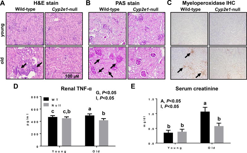
Histological and biochemical changes in aged WT compared to other groups. Aging induced profound histopathological changes in kidneys, as evidenced by increased inflammatory cell foci, tubular cytoplasmic vacuolation, tubular degeneration, glomerulonephropathy, and kidney fibrosis in old WT mice, which were significantly decreased in aged Cyp2e1-null mice, as shown with: (A) H&E staining, (B) PAS staining, and (C) myeloperoxidase IHC (n=7/group). Black arrows represent (A) inflammatory foci, (B) glomerulonephropathy and tubular degeneration/regeneration, and (C) myeloperoxidase. The levels of (D) renal TNF-α, and (E) serum creatinine are shown (n=6–8/group). A, age; G, genotype; I, age-genotype interaction. Labeled means, which do not share a common letter, represent significant difference from each other. P < 0.05.

