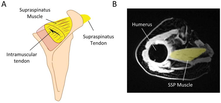
Fig. 1.
A) Schematic of supraspinatus (SSP) muscle and tendon. B) Volumetric masks were generated from both the fat and water magnetic resonance (MR) images (MR fat-image with masked SSP muscle is shown). Intramuscular fat fraction values were obtained from all masks based on the fat-water image datasets.

