
Figure 3.
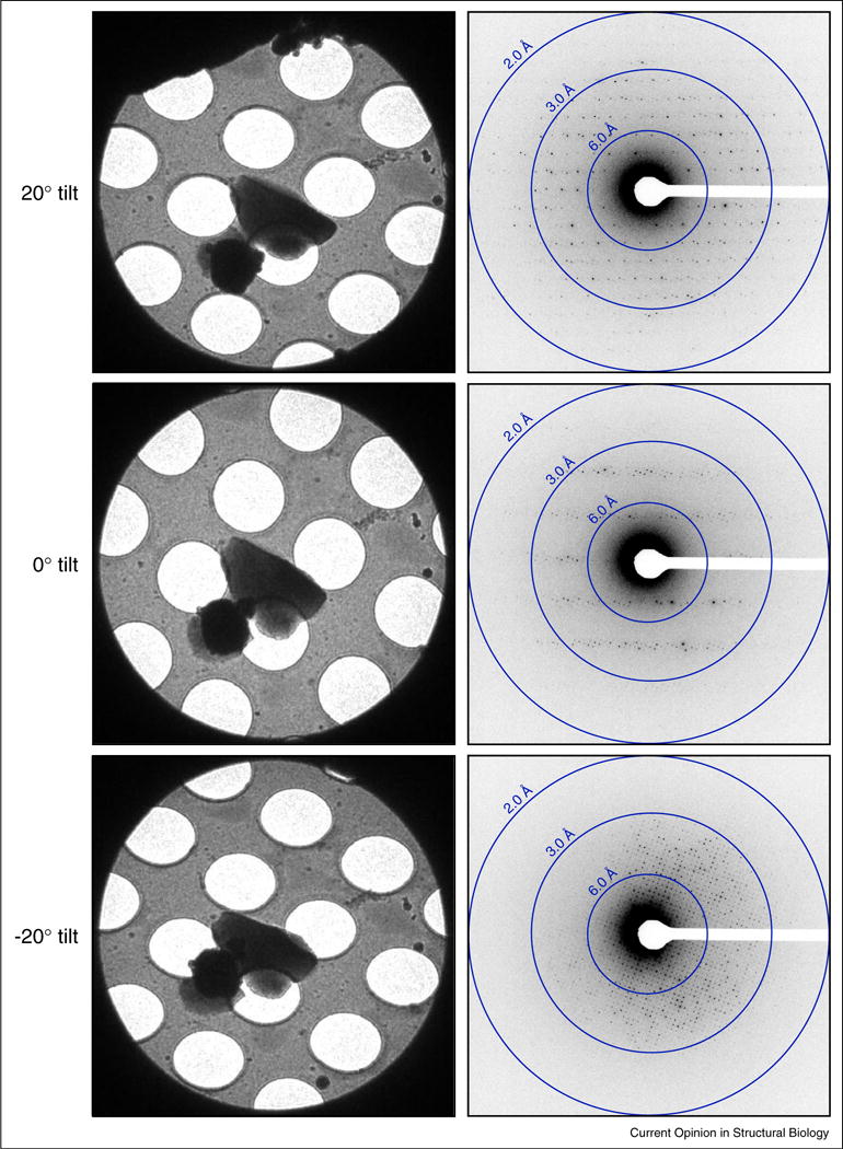
Examples of tilted diffraction data of Lysozyme collected by MicroED. Representative crystal images (left) and diffraction patterns (right) taken at tilts of 20°, 0°, and −20°. All images and diffraction patterns are from the same crystal. The MicroED diffraction data shows distinct reflections with a maximum resolution of 1.7 Å.