Figure 2.
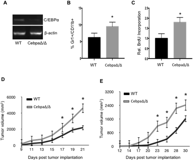
Tumor growth is accelerated in CebpaΔ/Δ mice. Gr-1+CD11b+ cells were purified from spleens of WT and CebpaΔ/Δ mice, RNA was isolated and C/EBPα expression was analyzed by semi-quantitative PCR (A). WT and C/EBPα CN mice were injected with 5 × 10⁵ 3LL (B–D) or B16 (E) tumor cells in the flank. After 21 days, spleens were isolated from the mice, processed into single-cell suspensions and stained with Gr-1 and CD11b fluorescent antibodies. The percentage of Gr-1+CD11b+ cells was analyzed by flow cytometry (B). 2 hours prior to sacrifice, the tumor bearing mice were injected with BrdU, and BrdU incorporation was measured in the Gr-1+CD11b+ cells by flow cytometry (C). Tumor dimensions were measured every 2–3 days with a caliper and tumor volume was calculated and plotted with time over time as indicated (D and E). *p < 0.05. n = 10 mice per group and repeated twice. The data are presented as mean with SD.

