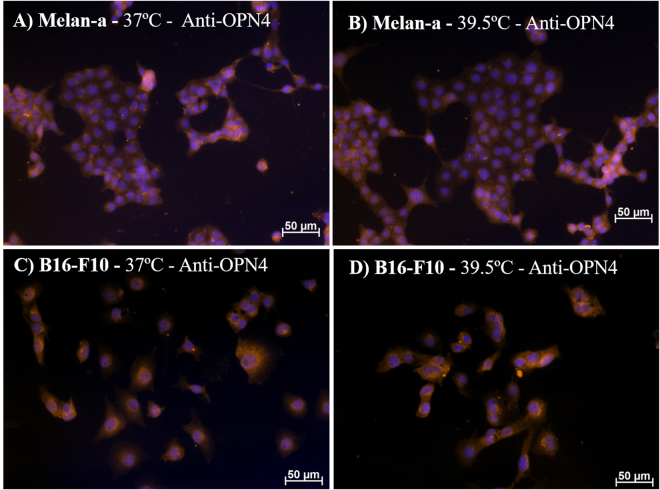
Figure 2.
Representative fields of melanopsin (OPN4) immunostaining in Melan-a (A,B) and B16-F10 (C,D) cells. Cells were kept in DD for 3 days and at the beginning of the 4th day, cells were divided into 2 groups: (1) Control group kept in constant dark and temperature (37 °C); (2) group in constant dark and exposed to 1 h heat stimulus (39.5 °C). Twenty-four hours later the medium was removed and the cells were fixed with 4% paraformaldehyde. DAPI stained nuclei in blue and OPN4 immunopositivity (1:500 antiserum), revealed with a Cy3-labeled secondary antibody, in orange. Photomicrographies were taken with Axiocam MRm camera (Zeiss) and pseudocolored with Axiovision software (Zeiss). Scale bar 50 μm (200x magnification).

