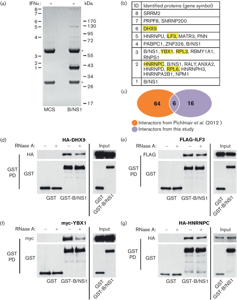
Fig. 2.
Identification of human proteins interacting with B/NS1. (a) Immunoprecipitation of V5-B/NS1 (or control) from IFN-stimulated cell lysates followed by SDS-PAGE and Coomassie blue staining. Protein bands specific for the B/NS1 lane are numbered. Asterisks indicate antibody heavy and light chain. Molecular weight markers (kDa) are indicated to the right. (b) Proteins identified by mass spectrometry performed on the gel slices cut from A. Each number (1–8) corresponds to the individual band analysed, and gene symbols of the proteins detected are listed. (c) Venn diagram of B/NS1 interactors identified in this study as compared with a previous screen performed by Pichlmair et al. The six B/NS1 interactors identified in both screens are highlighted in yellow in (b). (d–g) Western blot analysis of GST-pulldowns from 293 T-cell lysates co-transfected with the indicated plasmids for 48 h. Pull-downs were performed in the presence or absence of RNase A as indicated. Anti-tag antibodies were used to detect proteins of interest.

