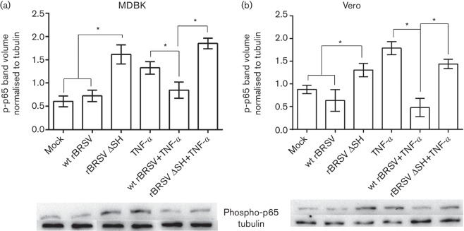
Fig. 1.
SH blocks phosphorylation of p65 in epithelial cells. MDBK (a) and Vero (b) cells were infected with recombinant virus (m.o.i. of 3) or mock-infected in the presence or absence of rTNF-α. Three hours post-infection, cell lysates were separated by sodium dodecyl sulfate polyacrylamide gel electrophoresis (SDS-PAGE) and levels of p65 phosphorylation determined by phospho-specific immunoblots. The graphs show phospho-p65 volumes normalized to tubulin volumes in the same sample. Columns indicate means of three replicates; error bars indicate standard deviation (sd); * indicates P<0.05. The blots shown are representative of three separate experiments.

