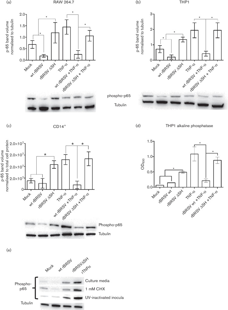
Fig. 2.
NF-κB p65 phosphorylation in APCs infected with rBRSV. RAW 264.7 (a), THP-1 (b) and bovine primary CD14+ cells (c) were infected with recombinant virus (m.o.i. of 3) or mock-infected in the presence or absence of rTNF-α. Three hours post-infection, cell lysates were separated by SDS-PAGE and levels of p65 phosphorylation determined by phospho-specific immunoblots. (a) and (b) Phospho-p65 volumes normalized to tubulin volumes in the same sample. (c) Phospho-p65 volumes normalized to total cell protein in each lane. (d) Alkaline phosphatase activity in culture supernatants from infected THP1-Blue NF-κB reporter cells. Columns indicate means of three replicates; error bars indicate standard deviation (sd); * indicates P<0.05. (e) NF-κB p65 phosphorylation in bovine primary CD14+ cells exposed for 3 h to Vero cell supernantant (mock), infected with wt BRSV, rBRSVΔSH (m.o.i. of 3), or treated with rTNF-α in: row 1, culture media; row 2, culture media containing 1 mM CHX; row 3, UV-inactivated inocula in culture media. All images are representative of three separate blots.

