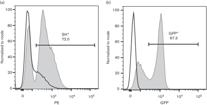
Fig. 3.
Expression of recombinant SH-V5/His6 in bovine CD14+. Primary bovine CD14+ cells were electroporated with pCDNA6 SH-V5/His6 (a) or pCDNA-GFP (b) or mock-electroporated (a and b). After 24 h in culture, cells were permeabilized and intracellular expression of SH-V5/His6 was detected using phycoerythrin (PE)-conjugated anti-V5; PE and GFP expression were measured by flow cytometry. Black histograms show autofluorescence of mock-electroporated cells stained with PE-conjugated anti-V5 (a) or autofluorescence alone (b); the grey-filled histogram shows fluorescence of electroporated cells stained with PE-conjugated anti-V5 (a) or GFP fluorescence (b). Figure representative of three experiments analysed in duplicate.

