Fig. 8.
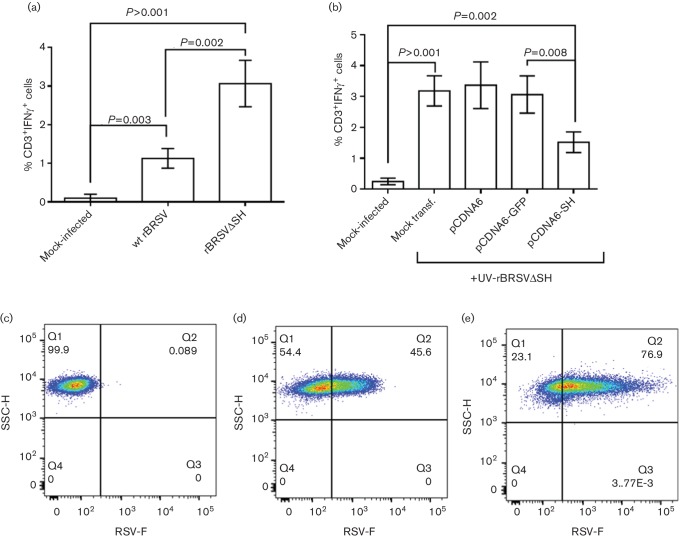
Expression of BRSV SH affects antigen presentation. Bovine primary CD14+ cells were (a) mock-infected or infected with wt rBRSV or rBRSVΔSH (both m.o.i. of 3) or (b) transfected with plasmids expressing SH or GFP and infected with UV-inactivated rBRSVΔSH. Activation of autologous T cells was detected by the expression of IFN-γ using flow cytometry. Bars indicate means of cells from four different animals analysed in duplicate. Error bars indicate standard error of the means. (c-e) Bovine primary CD14+ cells were mock-infected (c) or infected with an m.o.i. of 3 of wt BRSV (d) or rBRSVΔSH (e). Twenty-four hours later, the cells were fixed and stained with mouse anti-F. Dot plots are representative of cells from four different animals analysed in duplicate.

