Fig. 2.
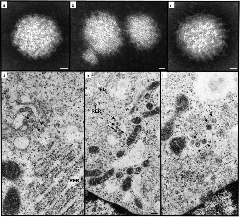
Transmission electron microscopy of WBV negatively stained particles (a, b, c) and infected Vero cells (d, e, f). (a) Virion of icosahedral-spherical shape, with an apparently amorphous envelope; (b) heterogeneity in particle size; (c) virus particle in which regularly spaced, envelope projections (right side) contrast with the fuzzy appearance of the envelope (left side); (d) transverse sections through characteristic tubular elements (arrows) forming within Golgi cisternal stacks, which are in close proximity to mitochondrial profiles and distended rough endoplasmic reticulum (RER); (e) developing virions (arrows) within a Golgi-derived vesicle, approaching the plasmalemma (PL) for exocytosis. Note the organelle arrangement with the nucleus (N) and numerous mitochondrial profiles in juxtaposition to the disrupted Golgi body; (f) mature virions (arrows) with glycoprotein envelope spikes, still within an endomembrane that appears continuous with those of the tangentially sectioned mitochondrial profiles. Scale bars: (a, b, c) 12 nm; (d, f) 100 nm; (e) 250 nm.

