
Figure 4.
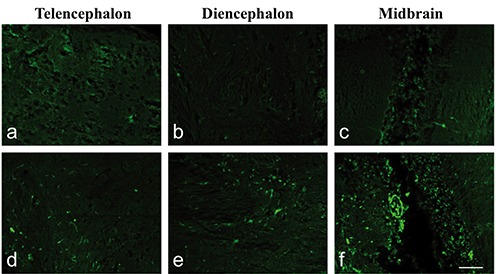
Fluoro-Jade B staining. Sagittal sections: a) telencephalon, b) diencephalon and c) midbrain in control adult fish; d) telencephalon, e) diencephalon and f ) midbrain of adults after 16 days treated with 9 µM of Cd. Note the increase in fluorescence in treated fish compared with the control. Fluorescence was especially present at the level of cell bodies and few nervous processes were stained. Scale bar: 50 µm.