
Fig. 3, A.
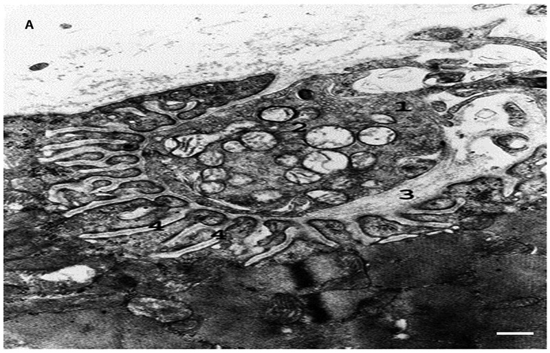
Electron micrograph of synapse of oxidative-glycolytic muscle fiber. 1. axon terminal; 2. synaptic vesicles; 3. synaptic cleft; 4. postsynaptic folds. Bar 0.5 μm
Official websites use .gov
A
.gov website belongs to an official
government organization in the United States.
Secure .gov websites use HTTPS
A lock (
) or https:// means you've safely
connected to the .gov website. Share sensitive
information only on official, secure websites.
Electron micrograph of synapse of oxidative-glycolytic muscle fiber. 1. axon terminal; 2. synaptic vesicles; 3. synaptic cleft; 4. postsynaptic folds. Bar 0.5 μm