
Figure 2.
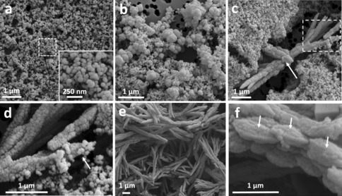
Time‐resolved SEM shows the progression of aragonite crystals, starting from amorphous calcium carbonate (ACC) particles. a) Sample collected after 1 minute of reaction, showing the presence of spherical ACC particles 75–100 nm in size. Inset: higher magnification of the area marked by the dotted square. b) Sample collected after 10 minutes of reaction, depicting 450–600 nm particles. c) Aragonite sheaf‐shaped crystals at 20 minutes of reaction. Arrow: calcite crystal growing at the expense of the surrounding aragonite. Dotted square: area shown in (d) at higher magnification. d) Higher magnification of the area marked by the dotted square in (c), showing spherical ACC particles attaching to the growing aragonite needle (arrow). e) Aragonite needles formed after 1 h of reaction. f) Higher magnification of one aragonite needle formed after 1 h of reaction, showing that they are composed of elongated particles of 900 nm–1 μm (arrows).