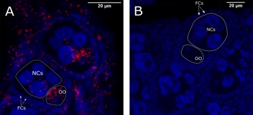
Figure 6.

Rickettsia localization in midge ovaries via FISH.
The combined z‐stack optical sections of infected C. impunctatus (A) and Rickettsia free C. nubeculosus (B) ovarioles stained with DAPI (blue) and an ATTO633‐labeled Rickettsia‐specific probe (red). FCs: follicle cells, NCs: nurse cells and OO: oocyte. [Color figure can be viewed at wileyonlinelibrary.com]
