
Figure 8.
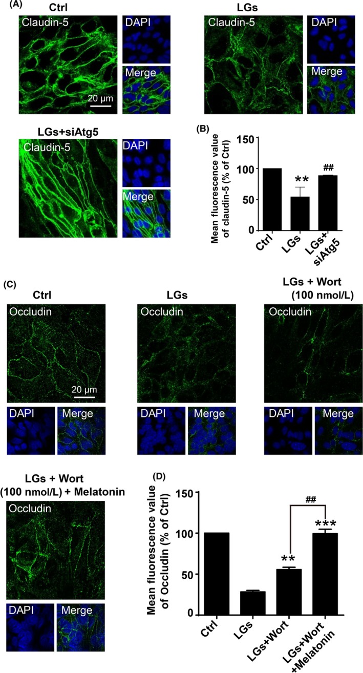
Inhibition of autophagy reduces tight junction injury during low‐glucose stress. (A) Representative immunofluorescence images of claudin‐5 staining of brain endothelial cells in the control and low‐glucose conditions. Cell cultures were transfected with siRNA‐Atg5, and Nuclei were stained with DAPI (blue). Scale bar, 20 μm. (B) Quantification of claudin‐5 expressions in (A). Data are expressed as the mean fluorescence intensity percentage ± SEM from 3 independent experiments. **P < .01 vs control; ## P < .01 vs low‐glucose‐treated cells. (C) Synergistically inhibitory effect of wortmannin and melatonin on degradation of occludin in brain endothelial cells upon low‐glucose insult. The cells were treatment with wortmannin (100 nmol/L) alone or co‐incubation with wortmannin (100 nmol/L) and melatonin (400 nmol/L) in low‐glucose cultures for 24 h. The nuclei were stained with DAPI (blue). Scale bar, 20 μm. Quantification of the fluorescence intensity of occludin is presented on (D). Florescence value is representative of 3 independent experiments expressed as mean ± SEM. **P < .01; ***P < .001 vs percentage of low‐glucose‐treated cells; ## P < .01 vs percentage of low‐glucose‐treated group with wortmannin alone