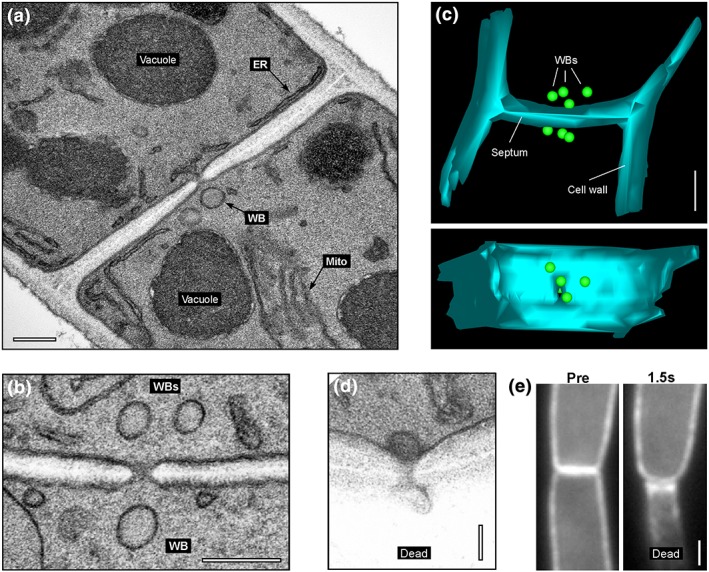
Figure 1.

Woronin bodies in Zymoseptoria tritici. (a) Electron micrograph showing a septum in Z. tritici. A single Woronin body (WB) is indicated. Scale bar represents 0.2 μm. (b) Electron micrograph showing a septum in Z. tritici. Several WBs surround the septal pore. Scale bar represents 0.2 μm. (c) A 3D reconstruction of serial sections through a septum of Z. tritici. Scale bar represents 0.5 μm. See also supplementary Movie S1 . (d) Electron micrograph of the septal pore of a wild‐type cell of strain IPO323 after wounding with quartz sand. A single WB has sealed the septal pore on the side of the intact cell. The injured cell has collapsed (dead cell). Scale bar represents 0.1 μm. (e) Behaviour of a septum, labelled with the plasma membrane marker eGFP‐Sso1, after laser‐induced rupture of the lower cell (indicated by “Dead”). The septum bends towards the collapsed cell, indicating a pressure gradient. Scale bar represents 1 μm. See also Movie S4
