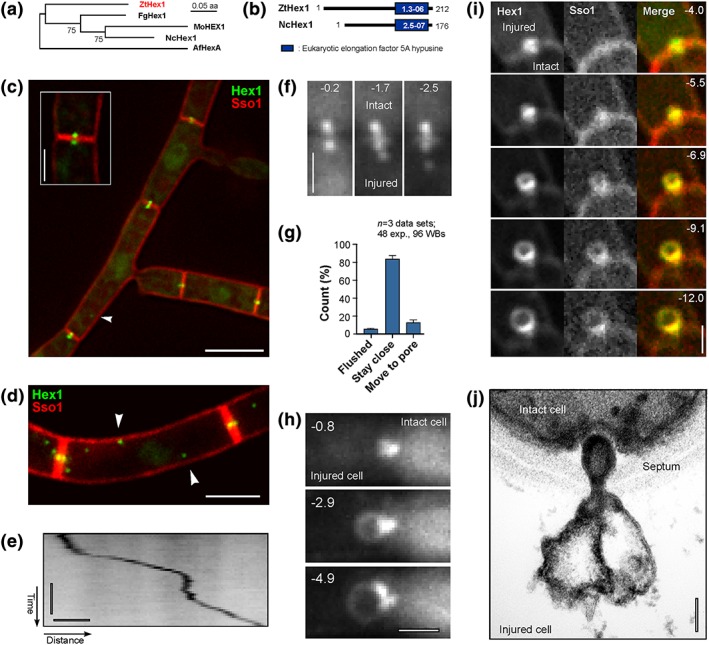
Figure 2.

Identification and live cell imaging of ZtHex1‐GFP. (a) Phylogenetic tree comparing the predicted amino acid sequence of fungal homologues of ZtHex1. NCBI accession numbers are as follows: Zymoseptoria tritici ZtHex1, XP 003854425.1; Magnaporthe oryzae MoHEX1, XP 003721069.1; Neurospora crassa NcHex1, EAA34471.1; Fusarium graminearum FgHex1, SCB65655.1; Aspergillus fumigatus AfHex, KMK59524.1. Maximum‐likelihood trees were generated using MEGA5.2. Bootstrap values from 500 rounds of calculation are indicated at branching points. Tree was generated in MEGA5.2; http://www.megasoftware.net/. (b) Comparison of the predicted domain structure of ZtHex1 from Z. tritici and NcHex1 from N. crassa. Error probabilities were determined in PFAM and are given in white numbers. (c) Z. tritici cells, coexpressing the Woronin body (WB) marker ZtHex1‐GFP and the red fluorescent plasma membrane protein mCherry‐Sso1. Strong ZtHex1‐GFP signals are concentrated on both sides of the septum (inset). In addition, nonmotile WBs of weaker fluorescent intensity locate in the cytoplasm (arrowhead). Scale bar represents 5 μm. (d) Maximum projection of a z‐axis stack of images, showing numerous cytoplasmic WBs (arrowhead). Scale bar represents 3 μm. (e) Contrast‐inverted kymograph showing directed motility of a cytoplasmic WB. Horizontal bar represents 2 s; vertical bar represents 1 μm. See also Movie S5 . (f) WB behaviour after laser wounding of Z. tritici cells. Immediately after injury, the cellular pressure drops in the wounded cell (lower half of images, indicated by “Dead”). The WB of the intact cell has plugged the septal pore, whereas WBs in the wounded cell remain stationary or move slightly away from the septum, whereas the cytoplasm bleeds out. Time after wounding is given in seconds. Scale bar represents 1 μm. See also Movies S6 and S7 . (g) Bar chart showing the behaviour of WBs in laser‐wounded cells. In most cases, the WBs in the ruptured cell stay associated with the septum. Mean ± standard error of the mean is shown; sample size n is 3 data sets, 48 experiments. (h) WB “ballooning” in a laser‐wounded cell of Z. tritici. After injury of the cell (left half of images, indicated by “Dead”), the WB of the intact cell plugs the pore. Within a few seconds, the WB balloons out, whereas the cytoplasm bleeds out of the ruptured cell. Time after wounding is given in seconds; scale bar represents 1 μm. See also Movie S8 . (i) Image series showing “ballooning” of ZtHex1‐eGFP and mCherry‐ZtSso1 after injury of a cell. The “balloon” contains the integral syntaxin ZtSso1, suggesting that the plasma membrane in the unwounded cell (indicated by “intact”) sealed after wounding and extends due to the pressure gradient into the wounded cell (indicated by “injured”). Time in seconds given in the upper right corner; scale bar represents 1 μm. See also Movie S9 . (j) Electron micrograph of the septal pore of a wild‐type cell after wounding with quartz sand. A WB has sealed the septal pore and formed a “balloon” into the injured cell (dead cell). Note that the membrane of the WB extends into the “bubble,” suggesting that the pressure gradient between the injured cell (dead cell) and the living cell (live cell) causes shape change of the organelle. Scale bar represents 0.1 μm
